“Early detection is the best protection” is a saying that has empowered people for decades to take charge of their health. It’s a phrase that rings true when it comes to just about any disease, but especially with breast cancer.
The 5-year survival rate for breast cancer is lower in advanced stages of the disease. Also, early detection remains one of the most effective strategies for preventing deaths from breast cancer.
Unfortunately, breast cancer is the second most common cancer found in women in this country (behind skin cancer), according to the U.S. Centers for Disease Control and Prevention.
While breast cancer can’t always be prevented, regular screenings and a healthy lifestyle have been shown to reduce a person’s risk. SummaCare sheds light on the different types of breast cancer screenings and which one(s) may be right for you.
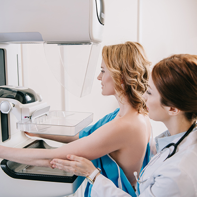
Since the 1960s, mammography (X-ray of the breast) has been the gold standard for diagnosing breast cancer in all stages. Luckily, as knowledge, research and equipment have advanced, so have screening technologies.
There are two types of mammograms available today: 2D and 3D mammograms.
A 2D mammogram captures two-dimensional flat images from the top and side of the breast, but 3D mammography is becoming more popular because it allows breast tissue to be examined layer-by-layer, in multiple views and with finer details. It’s especially useful for detecting cancer in females with dense breast tissue—resulting in earlier detection and decreasing the need for repeat testing for false positive findings.
If you are 40 or older, be sure to talk to your doctor about risk factors that determine how often you should have a screening mammogram. Females should start receiving annual mammograms no later than age 45.
For women at higher risk of breast cancer, or those with an abnormal mammogram, your provider may recommend one or more of the following tests:
Breast ultrasound uses sound waves and their echoes to make computer pictures of the inside of the breast. It’s useful for females with dense breast tissue to help providers better investigate abnormal findings identified on a mammogram or a palpable lump on examination.
A breast MRI uses magnets and radio waves after a contrast dye has been injected into a vein in the arm to take pictures of the breast. This test is used in conjunction with mammograms to screen women who are at higher risk for getting breast cancer.
The test takes between 45–60 minutes to complete and is an outpatient procedure. A provider order and preauthorization through your insurance is required.
This 15-minute, highly sensitive FAST Breast MRI uses contrast, magnets and radio waves to detect abnormal breast findings in women with dense breasts not seen on mammograms. The procedure does not involve any radiation exposure. A provider’s order is required for this screening.
SummaCare Professional